植體 - External 衛署醫器輸字第019542號

植體品牌-External植體圖示
原廠網站(外六角植體系統):
http://www.biohorizons.com/external.aspx
BIOHORIZONS 植體品牌 線上諮詢
*加LINE諮詢 : @hlb4651c
*加LINE連接 : https://goo.gl/k97R2E
BioHorizons美國植體品牌 植牙課程 植牙論文 植體產品諮詢

植體相關論文
相關學術文章:
Maintaining inter-implant crestal bone height via a combined platform-switched, Laser-Lok® implant/abutment system: A proof-of-principle canine study.
作者:M Nevins, ML Nevins, L Gobbato, HJ Lee, CW Wang, DM Kim.
出處:Int J Periodontics Restorative Dent , Volume 33, Number 3, 2013
摘要:
ABSTRACT
Interimplant papillae are critical to achieving esthetic implant-supported restorations in the maxillary esthetic zone. Stable papillary anatomy, however, depends upon a stable volume of underlying crestal bone for support. Multiple studies have documented a critical interimplant distance of 3 mm, under which crestal bone resorption occurs. The current preclinical proof-of-principle canine study examines a novel implant-abutment system design, combining platform switching with precisely configured laser-ablated abutment and implant microgrooves to maintain interimplant crestal bone at interimplant distances of 2 and 4 mm. Results of this initial preclinical study suggest that it is possible through precise implant/abutment design modifications to place adjacent implants at distances of 2 to 4 mm without inducing subpapillary crestal bone loss.

載入全文
相關學術文章:
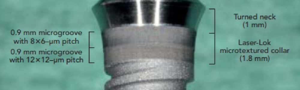
Radiographic analysis of crestal bone levels around Laser-Lok collar dental implants.
作者:CA Shapoff, B Lahey, PA Wasserlauf, DM Kim.
出處:Int J Periodontics Restorative Dent. 2010;30:129-137
摘要:
INTRODUCTION
This retrospective radiographic study was organized to evaluate the clinical efficacy of implants with Laser-Lok microtexturing (8- and 12-μm grooves). A physical attachment of connective tissue fibers to the Laser-Lok microtexturing on the implant collar has been previously demonstrated using human histology, polarized light microscopy, and scanning electron microscopy. This analysis of 49 implants demonstrated a mean crestal bone loss of 0.44 mm at 2 years postrestoration and 0.46 mm at 3 years. All bone loss was contained within the height of the collar, and no bone loss was evident to the level of the implant threads. The radiographic evaluation of the clinical application of this implant supports previous findings that establishing a biologic seal of connective tissue fibers around a dental implant may be clinically relevant.
載入全文
 |
|
|
| 線上諮詢 | 食藥署核准仿單 |
植體相關資訊


植體相關資訊
|
Tapered Internal |
Tapered PLUS  |
Internal  |
|
External
|
Laser-Lok3.0 |
|
|
|


植體特色
相關產品