
利用X光片評估有Platform-Switched及雷射微溝槽的植體在狹小空間的臨床表現。
A prospective clinical and radiographic assessment of platform-switched laser-microchannel implant placed in limited interimplant spaces.;
論文出處:
The International Journal of Periodontics & Restorative Dentistry Volume 37, Number 1, 2017
The International Journal of Periodontics & Restorative Dentistry Volume 37, Number 1, 2017
INTRODUCTION介紹
The objective of the present prospective clinical study was to investigate the hard and soft tissue result when implants are placed <3mm apart to emulate the interproximal space between teeth in the esthetic zone.
本臨床研究的目的是評估植體植入在前牙區小於3mm的空間下軟硬組織的狀況。
本臨床研究的目的是評估植體植入在前牙區小於3mm的空間下軟硬組織的狀況。
MATERIALS AND METHODS材料和方法
A total of 38 implants were placed in 18 patients and evaluated at 1 year or longer after restoration. Each patient presented with a localized edentulous ridge site requiring two dental implants placed 2 to 3 mm apart. This situation allowed evaluation of soft and hard tissue behavior.
18名病患共植入38支植體,並於贗復至少一年以上的時間後進行評估。每名患者的齒槽骨皆具有局部缺牙的條件,兩支植體植入的間距需有2-3mm的空間,並進行軟硬組織的評估。


RESULTS結果
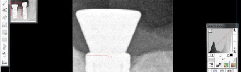
After 1 year of loading, the inter-proximal bone appeared to be at the level of the implant abutment junction for most of the implants. No significant osseous dieback was seen around 31 implants, but 4 implants lost bone to the first thread and 1 implant to the second thread.
受力一年後,大部份植入的植體與近端齒槽骨間距落在植體與支台齒接合點的水平處。在31支植體周圍無顯著的齒槽骨萎縮,但有4支植體齒槽骨流失到第一螺紋處,1支植體齒槽骨流失到第二螺紋處。

CONCLUSIONS結論
Radiographic and photographic evidence provide an optimistic outlook for this system. Most cases demonstrated intact interdental papillae and no loss of bone apical to the collar of the implant. No recession was evident in any of the implants. There was a lack of total interproximal soft tissue papillae in 3 of the 38 cases.
X光片的證實對此系統提供了一個樂觀的前景。大部分的案例表現出完整的papillae,且植體頸部並無齒槽骨流失,也沒有軟組織明顯萎縮的狀況。38個案例中僅有3例缺乏papillae。


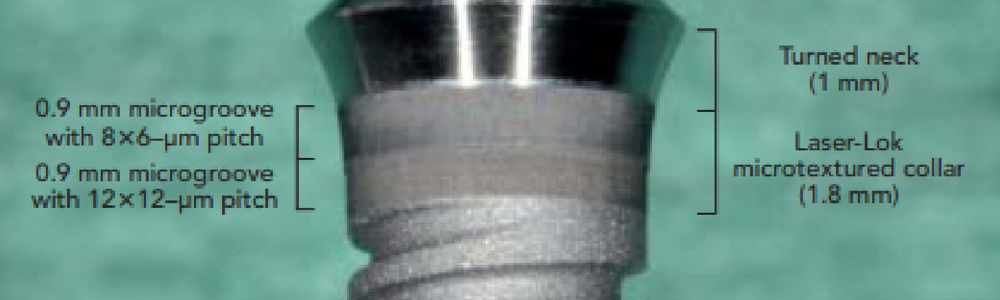
植 體 特 色
植體特色


©2026 BioHorizons美國植體品牌 - 在台16年植牙植體品牌、各國植牙成功案例、植牙體品牌、 聯雄健康事業股份有限公司 | Designed by Hurricane Media
